38 knee joint with labels
Knee Joint Label Diagram | Quizlet Start studying Knee Joint Label. Learn vocabulary, terms, and more with flashcards, games, and other study tools. A Labeled Diagram of the Knee With an Insight into Its ... Labeled Diagram of the Knee Joint Knee joint is one of the most important hinge joints of our body. Its complexity and its efficiency is the best example of God's creation. The anatomy of the knee consists of bones, muscles, nerves, cartilages, tendons and ligaments. All these parts combine and work together.
A Diagrammatic Explanation of the Parts of the Human Knee ... Knee actually consists of three bones - femur, tibia and patella. Femur is the thigh bone, tibia is the shin bone and patella is the small cap like structure which rests on the other two bones. Femur is considered as the largest bone in the human body. The femur and the tibia meets at the tibiofemoral joint and patella rests on top of this joint.
Knee joint with labels
The Knee Joint - Articulations - Movements - Injuries ... The knee joint is a hinge type synovial joint, which mainly allows for flexion and extension (and a small degree of medial and lateral rotation). It is formed by articulations between the patella, femur and tibia. In this article, we shall examine the anatomy of the knee joint - its articulating surfaces, ligaments and neurovascular supply. Knee Joint Picture Image on MedicineNet.com The knee functions to allow movement of the leg and is critical to normal walking. The knee flexes normally to a maximum of 135 degrees and extends to 0 degrees. The bursae, or fluid-filled sacs, serve as gliding surfaces for the tendons to reduce the force of friction as these tendons move. The knee is a weight-bearing joint. Knee (Human Anatomy): Function, Parts, Conditions, Treatments The knee is one of the largest and most complex joints in the body. The knee joins the thigh bone (femur) to the shin bone (tibia). The smaller bone that runs alongside the tibia (fibula) and the ...
Knee joint with labels. Label The Structures Of The Knee Joint - Solved Procedure ... Label the structures of the knee joint (anterior view) by clicking and dragging the labels to the correct location. To deepen the articular surface of the tibia, thus increasing . The knee joint has three parts. The knee joint keeps these bones in place. The joint head on the femur has . Knee joint: anatomy, ligaments and movements | Kenhub The tibiofemoral joint Medial condyle of femur Condylus medialis femoris 1/7 The tibiofemoral joint is an articulation between the lateral and medial condyles of the distal end of the femur and the tibial plateaus, both of which are covered by a thick layer of hyaline cartilage . Knee Anatomical Models | Knee Joint Models Knee Models. Because the knee supports almost all of one's body weight, this joint is highly susceptible to injury. With the help of knee models, however, it is easy to see how the three main components, the femur, patella, and tibia, work together, and how they can buckle under stressful situations. Knee education models are useful classroom ... 3D Knee Joint Model *Finished Product* - YouTube Finally completed my knee joint model with labels of all the key ligaments, muscles, tendons, and bursae. Let me know what you think, I spent a lot of time ...
Knee Joint Label Flashcards - Quizlet Knee Joint Label STUDY Flashcards Learn Write Spell Test PLAY Match Gravity Created by LaLaKub91 Terms in this set (10) femur What is A? lateral collateral ligament what is d? lateral meniscus what is e? fibula what is g? tibia what is h? posterior cruciate ligament What is j? anterior cruciate ligament what is k? medial meniscus what is l? Knee Joint - Anatomy Pictures and Information The knee, also known as the tibiofemoral joint, is a synovial hinge joint formed between three bones: the femur, tibia, and patella. Two rounded, convex processes (known as condyles) on the distal end of the femur meet two rounded, concave condyles at the proximal end of the tibia. Continue Scrolling To Read More Below... Additional Resources Alila Medical Media | Knee joint, basic labels | Medical ... Human knee joint diagram showing joint cavity, capsule, all cartilage. - Alila Medical Media Amazon.com: anatomical model knee Axis Scientific Functional Knee Model - Anatomically Correct Knee Joint with Life Like Ligaments That Can Show Movement, Includes Base, Detailed Full Color Product Manual, Worry Free 3 Year Warranty 22 $49 99 Get it as soon as Wed, Apr 13 FREE Shipping by Amazon
Knee x-ray - labeling questions | Radiology Case ... Normal X-ray Knee - Frontal (with labels) Annotated image Frontal Knee Frontal 1. Femoral shaft 2. Patella 3. Base of patella 4. Apex of patella 5. Adductor tubercle of femur 6. Medial epicondyle of femur 7. Medial condyle of femur 8. Lateral epicondyle of femur 9. Lateral condyle of femur 10. Groove for popliteus 11. Intercondylar fossa 12. Knee Diagram - Pro Knee Pain Relief The knee is a complex joint that has many functions. It bends back and forth and twists minimally from side to side. The knee connects the thigh bone (femur located in the upper leg) to the shinebone (tibia located in the lower leg.The calf bone (fibula located in the lower leg) connects to the joint, but is not directly affected by the hinge joint action. Knee (Human Anatomy): Function, Parts, Conditions, Treatments The knee is one of the largest and most complex joints in the body. The knee joins the thigh bone (femur) to the shin bone (tibia). The smaller bone that runs alongside the tibia (fibula) and the ... Knee Joint Picture Image on MedicineNet.com The knee functions to allow movement of the leg and is critical to normal walking. The knee flexes normally to a maximum of 135 degrees and extends to 0 degrees. The bursae, or fluid-filled sacs, serve as gliding surfaces for the tendons to reduce the force of friction as these tendons move. The knee is a weight-bearing joint.
Knee Joints
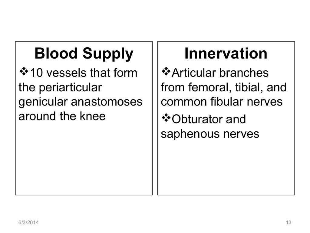
The Knee Joint - Articulations - Movements - Injuries ... The knee joint is a hinge type synovial joint, which mainly allows for flexion and extension (and a small degree of medial and lateral rotation). It is formed by articulations between the patella, femur and tibia. In this article, we shall examine the anatomy of the knee joint - its articulating surfaces, ligaments and neurovascular supply.

Anatomy of knee joint Mousepad | Zazzle

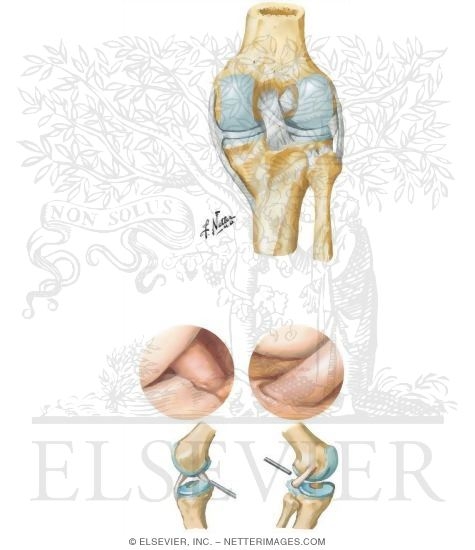
Medical Education resources and tools | Elsevier
![09 [chapter 9 joints]](https://image.slidesharecdn.com/09chapter9joints-170828041032/95/09-chapter-9-joints-40-638.jpg?cb=1503893470)
09 [chapter 9 joints]

Knee Joint (Lateral)
Anatomy of the knee joint
Post a Comment for "38 knee joint with labels"